Teeth and bones of the European sabre-toothed cat
Paläon Schöningen - Germany
Contact us
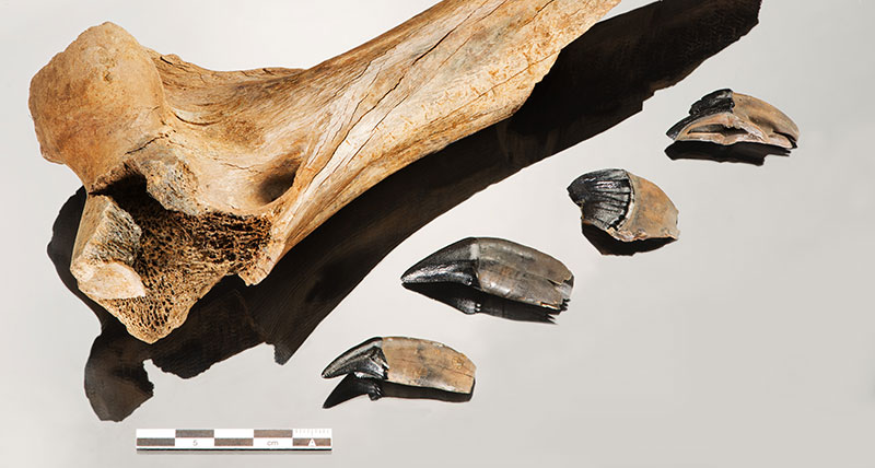
The curved canine tooth serves as namesake — and its length of more than 10 cm indicates at the same time that you cannot compare the extinct European sabre-tooth cat with today‘s domestic cat: The bone and tooth remains found in Schöningen (Germany) belonged to a young animal with a shoulder height of approximately 1.1 meters and about 200 kilogramms weight. Nowadays, this physique corresponds to the body of a lion or a tiger!
Objective and measuring object
The sabre-toothed cat (Homotherium latidens) was widely spread in the Early and Middle Pleistocene. Therefore, you often find remains of these animals at archaeological excavations, but complete skeleton finds are rare. The finds from Schöningen are approximately 300,000 years old, but still they impressively demonstrate how dangerous its former possessor had been. Equally dangerous for our ancestor, the Homo heidelbergensis, because the bone and tooth remains of the excavations at Schöningen prove that our ancestors and the carnivore had lived at the same time in the area of the Schöningen lakeside — and with the utmost probability, they had even met each other.
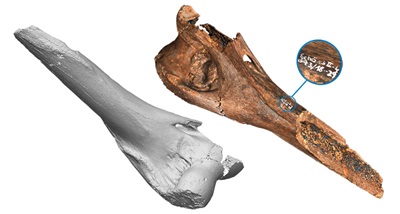
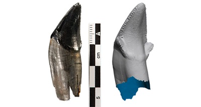
The preservation in humus soil blackened the shiny dental enamel, a typical appearance of animal remains at this archaeological site. A thorough investigation of all details of the irregular formed bone and tooth shapes with a size of only a few centimeters requires highly precise three-dimensional captures — this kind of digitization project calls for the SmartScan.
Measuring system and setup
Tactile measuring techniques only partially suit for delicate fossil objects that might be damaged by the probing. For the contact-free digitization of the finds, the SmartScan is applied. Thanks to its modular configuration using black-and-white or color cameras in varying resolutions, this AICON white-light scanner allows for exact adjustment to the requirements of users and their projects.
The three-dimensional capturing is carried out with two 5 Megapixel color cameras. For the teeth, a field of view size of 60 mm is applied and a size of 125 mm for the bone. These configurations of the fringe-projection scanner are especially suitable for the detailed capturing of artefacts with a size of approximately 20 cm to less than 5 cm. In addition, a turntable facilitates the complete scanning of the finds.
Workflow
Capturing the black shiny surfaces of the dental enamel is a particular challenge to a scanner, especially as no matting spray is used. The system calibration is followed by the 3D measurement with the SmartScan. The digitization process consists of several measuring sequences. Then the AICON software OptoCat merges the single pictures into the find‘s complete digital representation including all its features down to the finest detail. The precise 3D model of the tooth serves as the ideal basis for detailed research as well as for documentation and archiving.
The SmartScan‘s color cameras capture the find‘s form and its texture at the same time. This texture can be mapped in high resolution onto the digital object data — an easy working step for the Texture Mapping module of the OptoCat software.
Result
The SmartScan captures the very sensitive paleontological finds in a contact-free, fast, and highly precise way. Using the digital representation for research protects the precious object. The 3D scan data even allow for more detailed analyses of the dental enamel‘s surface than the original object.
The texture mapping can be carried out both by using the internal imagery of the SmartScan as well as using images taken with any kind of external camera. Working with external digital data not only allows for a variety of possibilities of texture capturing but also provides a large degree of freedom with regard to pixel resolution, professional illumination and raw data processing with color management. Furthermore, it is possible to complement the 3D data generated by a monochrome scanning sensor with color imagery, or even to work with multispectral data (UV, IR, etc.).
With the OptoCat Texture Mapping module, the 3D model gets an improved color reproduction, a clearer contrast and a higher texture resolution — and therefore reaches a much more realistic representation of the original object than the scanner cameras‘ mere vertex colors.
We would like to thank the Paläon Schöningen and the Landesmuseum (State Museum) Hannover for their kind support in providing information and pictures used to compile this application report.
Some of the products covered in this case study have since been discontinued.
Please visit this page for details of our current options in this product category.

