Analisi multibody di un chiodo endomidollare auto-bloccante per il trattamento di fratture del femore
It was assessed that the diaphyseal fractures of the femur, caused by violent trauma, occurred with a fracture frequency of every 10,000 people. The categories affected are young people under the age of 25 and the elderly over 65 and the causes can be many.
The use of appropriate surgical techniques and not, combined with new technologies and devices, is a solution that reduces the costs of the national health system by reducing the time of recovery of the patient and improving at the same time his general state of health. The self-locking intramedullary nails appear as devices which, with respect to conventional endomidullary nails, aim to reduce the injuries inflicted on the patient during the implantation operation of the fixing device. From a functional point of view, the device must be able to guarantee the maintenance of an appropriate alignment of the two femoral bone segments during the fracture healing process and to allow the transfer of an external load from one bone segment to the other avoiding that the collapse of the fracture.
The aim of this work was to examine a new idea of a self-locking elastic endomidullary nail, analyzing its dynamic behavior in the implant configuration. In particular, the system resulting from the coupling between the intramedullary nail and the femoral bone segments was analyzed through a multibody approach that allowed to obtain information on displacements, deformations and forces exchanged between the different components of the system.
The device can assume a closed or open configuration, the first is reached to correctly insert the nail inside the bone, while the second one is obtained at the end of the stem opening which, coming into contact with the medullary canal, allows to keep the bone fixed in the correct position. At this point, the bone-implant system has been subjected to bending, torsional and compression loads; in particular for the compression test a sine force was applied at 750N amplitude, while for the flexion 175N, both lasting 2 seconds and with a frequency of 0.25 Hz. For the torsion a sinusoidal torque with a width of 500N * mm was applied. the duration of 6 seconds, also in this case at 0.25 Hz frequency.
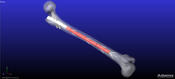
The simulations, using MSC Adams multibody code and FE Part technology (see Figures) , foresee the succession of a closing and opening test of the device inside the medullary canal, followed by one of the three torsion, flexion or compression tests.
The closure of the device highlights the proportionality between the force necessary to confine the stems inside the mask and the stem-bone interaction force, in order to have an increase in the second one necessarily increases the former.
The opening phase, on the other hand, shows problems concerning above all the contact between the stems and the medullary canal that occurs in an instant that does not coincide with the end of the opening but first, in a time that depends on the stem geometry and the medullary canal section.
During the flexion test the main problems encountered were in the distal segment of bone where there was a flexion of 4.5 ° with respect to 0.6 ° of the proximal one, where this was limited by the presence of the stub.
In the torsion test the device was not able to fully compensate the external torque, obtaining an overall rotation of the distal segment of the bone with respect to the initial position of the bone of almost 180 °.
However, the compression test is the most problematic, where the limits of this device are highlighted. We have seen from the simulations how even minimal compressive forces (10N), cause a mobility of the device. Having tried an alternative geometry that guaranteed with the distal stems a greater contact force does not lead to good results, after having shown also in this circumstance the failure of the device for loads of the order of 60N.
It is therefore not possible to use this device in patients who need mobility since a few days after the operation since it would probably fail, but it may be intended for bedridden patients who do not need to charge the device. A possible improvement to be made to this study would be to resort to a change in the design part of the geometry, in order to overcome the limits found by this specific configuration. Moreover, the bone-implant contact could be studied in more detail, modeling also the spongiosa component, in order to find a solution as close as possible to the real situation.